
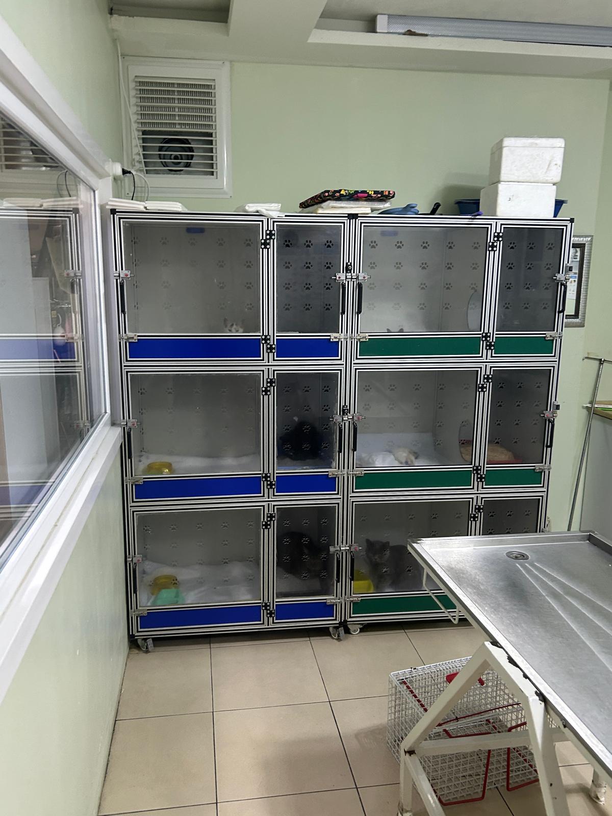
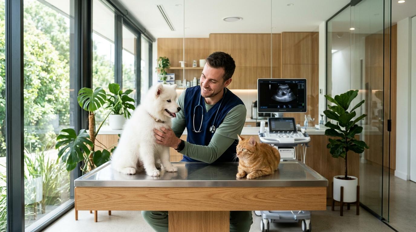
In-House Laboratory and Result Flow
At our in-house laboratory in Tatlıkuyu, Gebze (serving Darıca, Çayırova, Dilovası and Kocaeli), a complete blood count guides the assessment of anaemia, infection and clotting disorders through red cell, white cell and platelet values. The biochemistry panel covers liver enzymes, renal parameters, glucose, protein and electrolytes, and is decisive when calculating treatment doses.
Urinalysis supports renal and urinary tract assessment as well as diabetes monitoring; a first morning sample is preferred and is read together with sediment microscopy. Rapid antigen tests such as parvovirus or FeLV / FIV allow isolation and treatment to start the same day.
- CBC and biochemistry results reviewed during the same visit
- External laboratory confirmation for equivocal cases
- Trend comparison against previous results in chronic patients
Preparation Standards for Radiography and Ultrasound
Plain radiography is first-line for bone, joint and thoracic assessment, with a four-view thoracic series as standard. Abdominal ultrasound depends on tissue contrast, so an 8 to 12 hour fast and a full bladder where possible markedly improve image quality.
Related Clinical Procedures & Health Guides
| Method | Clinical Purpose | Patient Preparation | Turnaround |
|---|---|---|---|
| Complete blood count (CBC) | Anaemia, infection, inflammation monitoring | 8 hour fast recommended | Same visit |
| Serum biochemistry | Liver, kidney, glucose, electrolytes | 8 – 12 hour fast | Same visit |
| Urinalysis | Renal, urinary tract, diabetes | First morning sample preferred | Same visit |
| Plain radiography | Bone, joint, thorax | No anaesthesia required | Same visit |
| Abdominal ultrasound | Organ parenchyma, effusion, mass | 8 – 12 hour fast | Same visit |
| Rapid antigen test | Parvovirus, FeLV / FIV screening | No extra preparation | 15 minutes |
Clinical Pathway
- The examination defines the clinical question and the priority test list.
- Sample collection and patient preparation for imaging are completed.
- Results are combined with physical examination findings by the veterinarian.
- Diagnosis, treatment plan and recheck schedule are explained to the owner.
Frequently Asked Questions
Is fasting required for an ultrasound?
Yes. Because stomach and intestinal gas reduce image quality, an 8 to 12 hour fast is usually recommended. Water is not restricted, and a full bladder makes the assessment easier.
Does radiography harm the animal?
Plain radiography needs no anaesthesia and is a safe examination at diagnostic doses. Protective equipment for staff and correct patient positioning prevent unnecessary repeat exposures.
How often will repeat blood work be needed?
That depends on disease stage and response to treatment. Chronic cases are monitored at intervals between two and twelve weeks, with each result compared against the previous trend.
The information on this page is for general guidance and does not replace a physical examination.